How the Human Brain Maps Uncertainty and Prepares for the Unexpected
The Biology of Foresight: Uncovering How Our Memory Hub Primes Us for Surprises.
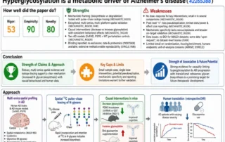
Tessa analyzed a recent study on how the brain predicts and manages uncertainty. The study produced an overall Trustworthiness Score of 83, placing it in our top 5% of research.
According to predictive coding formulations, the brain manages this by continuously generating top-down predictions about upcoming sensory inputs. When a mismatch occurs between what is predicted and what is observed, a bottom-up prediction error is returned to update higher-level representations. Crucially, these prediction errors are weighted by the level of uncertainty (their precision) associated with the context, balancing prior beliefs against sensory evidence.
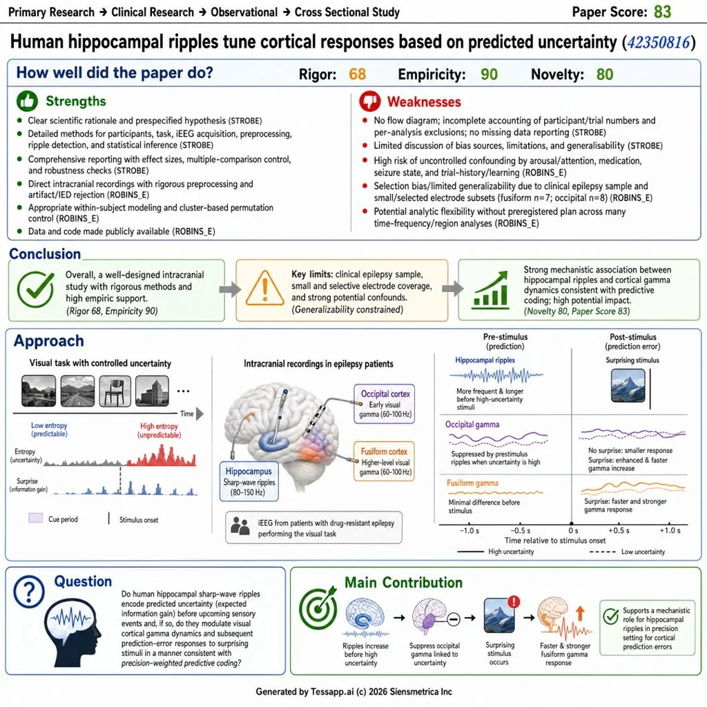
A recent study published in Nature Neuroscience by Frank et al. (2026), titled “Human hippocampal ripples tune cortical responses based on predicted uncertainty,” explores the precise neural mechanisms backing this phenomenon. Specifically, the researchers investigated whether human hippocampal sharp-wave ripples (SWRs), historically studied for their role in memory consolidation and replay, serve a prospective role in encoding uncertainty and modulating cortical dynamics before an event even occurs.
The Experimental Framework
To study how the brain maps the unexpected, the research team looked directly inside the human brain. They analyzed direct intracranial brain recordings (iEEG) from 17 surgical epilepsy patients.
While patients performed a simple visual matching task on a screen, researchers altered the mathematical probability of the images being shown. This allowed them to cleanly separate two distinct types of information data points:
- Entropy (The “Before” Metric): This scores the overall uncertainty or unpredictability of an environment before an event occurs.
- Surprise (The “After” Metric): This scores the unexpectedness of a specific event after it happens, measuring how severely your predictions were violated.
Why this matters: In daily life, surprise and uncertainty usually happen at the same time. By separating them mathematically, the researchers could pinpoint exactly how the brain prepares for an event versus how it reacts to it.
What the Data Reveals
The study uncovered a perfectly timed, highly synchronized dialogue between the memory center of the brain (the hippocampus) and the visual cortex:
Step 1: The Brain Spots Uncertainty. When an environment becomes highly unpredictable, the hippocampus immediately increases the frequency and duration of its “sharp-wave ripples”—bursts of rapid electrical activity—before any image even appears.
Step 2: The Hub Primes the Visual System. These prestimulus ripples act as a strategic filtering command. They travel to the visual cortex and suppress raw, unweighted uncertainty noise (gamma-band activity).
Step 3: An Accelerated Reaction to Surprise. By suppressing that background noise early, the ripples shift the higher-level visual processing stream into a perfectly balanced, receptive state. Because the brain invested resources upfront to map the uncertainty, its subsequent response to a surprising image is 200 milliseconds faster and significantly stronger.
The Direction of Information Flow: Granger causality analysis confirmed a strict temporal split in communication paths. Top-down directives flow out from the hippocampus to prime the cortex before an event. Bottom-up prediction errors flash back from the visual cortex to the hippocampus after a surprise occurs.
Implications
The implications of these findings extend far beyond the laboratory, offering a potential biological blueprint for several real-world applications. By understanding how the brain actively manages an “entropy budget,” we can develop more efficient artificial intelligence architectures that dynamically adjust processing based on uncertainty.
Clinically, this ripple-mediated mechanism could serve as a vital biomarker for diagnosing neurological and psychiatric conditions involving miscalibrated prediction errors. Furthermore, in high-stakes fields like aviation or emergency response, these insights could inform training protocols that condition trainees to better “prime” their neural responses to unpredictability, potentially enhancing reaction times and decision-making accuracy in complex environments.
Tessa Assessment Insights
Overall T-Score: 83/100 (Green 🟢)
We ran the paper through our evaluation platform, Tessa, which analyzed this paper across multiple reporting and methodological domains, yielding an overall Trustworthiness Score (TScore) of 83.
Analytical Breakdowns:
- Theoretical vs. Experimental (90/100): Strongly empirical. The study relies heavily on direct human intracranial data, robust signal processing, and convergent mixed-effects modeling rather than abstract theory development.
- Known to Novel Method (80/100): While sharp-wave ripples and predictive coding frameworks are well established independently, linking prospective human ripple dynamics to precision-weighted cortical processing in the absence of memory demands represents a significant and innovative step forward.
- Weak to Rigorous (68/100): Methodological execution is highly transparent, featuring stringent interictal epileptiform discharge (IED) rejection and analytical replication using an alternative ripple-detection algorithm.
Methodological and Reporting Gaps
A deep dive using standardized protocols highlighted specific execution profiles:
- Reporting Completeness (STROBE Checklist – 90%): The study provides excellent detailing of behavioral tasks, electrode localization, and time-frequency statistics. Gaps exist in the standard observational terminology layout in the abstract, the omission of a participant/trial attrition flow diagram, and incomplete clinical data regarding exact baseline medication profiles.
- Risk of Bias (ROBINS-E): Rated as High Risk. This is inherent to the clinical nature of intracranial electrophysiology studies. The participant pool is restricted to a highly selective surgical epilepsy cohort, and sample sizes for specific cortical regions are small (occipital $n=8$, fusiform $n=7$). Furthermore, observational within-subject designs face challenges from unmodeled time-varying confounders like sudden fluctuations in patient attention, fatigue, or residual sub-clinical seizure activity.
Summary Data
- Verified References: 77 out of 77 (100% verified, with a 6.6/10 average context relevance score).
- Data/Code Status: Open and accessible via GitHub.
To view the complete metrics, including researcher-institution profiles, citation timelines, and a structural breakdown of the statistical domains, read our Full Tessa Summary Report.
Share This →
About the Author: Sage Osterfeld
Research Spotlight

Recent Posts


